
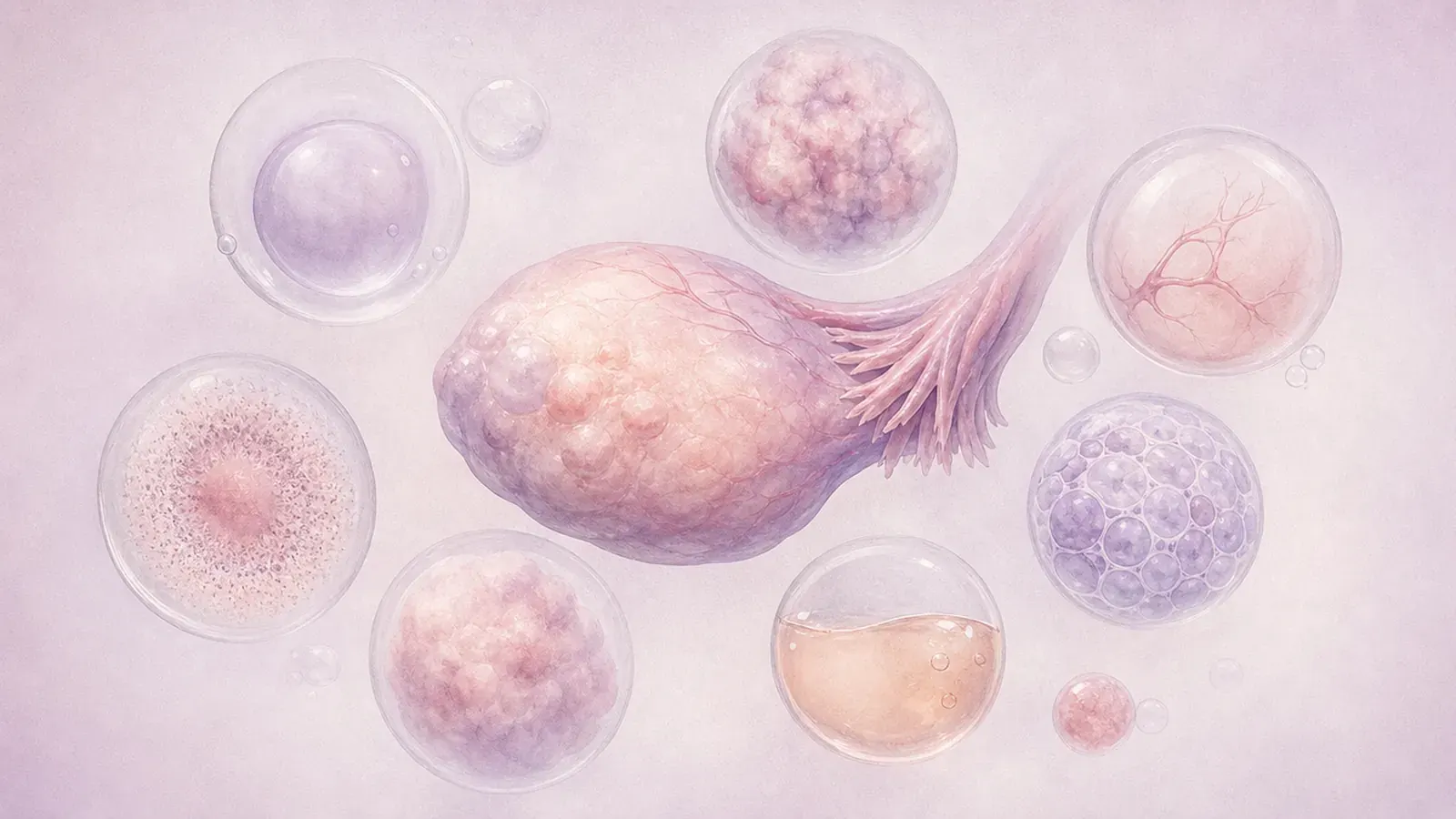
Seeing "ovarian cyst" on an ultrasound report often causes worry. Yet a cyst simply means a sac filled with fluid or semi-fluid content, and the great majority of ovarian cysts are benign. Whether a cyst should be watched, treated with medicines or removed depends largely on its type. This article looks at the common types one by one. For the general assessment, symptoms and treatment options, see our page on ovarian cysts.
Functional cysts: part of the cycle
Functional cysts are not a disease but a natural by-product of ovulation. They can occur at any age while you have periods and are not expected after the menopause. Most cause no symptoms and disappear on their own within a few weeks to a few cycles.
Follicular cyst
Each month a sac carrying an egg (follicle) grows in the ovary and bursts at ovulation. If the follicle does not burst and keeps filling with fluid, a follicular cyst forms. On ultrasound it looks like a round, thin-walled sac of clear fluid, described as a "simple cyst".
Corpus luteum cyst
After ovulation the empty follicle turns into the corpus luteum ("yellow body"), which produces progesterone. If fluid collects inside it, a corpus luteum cyst forms. Its wall is thicker, and colour Doppler ultrasound may show a ring of blood flow around it. It is also common in early pregnancy, where it makes hormones that support the pregnancy, and usually shrinks by itself as the pregnancy progresses.
Haemorrhagic cyst
This forms when there is bleeding into a follicular or corpus luteum cyst and can cause sudden one-sided pelvic pain. On ultrasound it looks like a cyst with a net- or lace-like pattern inside, or a denser area caused by a clot. Most are absorbed within a few weeks; your doctor usually confirms that the cyst has gone with a follow-up scan in a later cycle.
For functional cysts, the first approach is usually observation. Contraceptive pills have been shown not to speed up the disappearance of an existing functional cyst, although a doctor may suggest them for some women who get cysts often, to suppress ovulation and reduce new cysts. If a cyst does not disappear in the expected time, grows or changes in appearance, another type of cyst is considered.
Endometrioma ("chocolate cyst")
An endometrioma is a cyst in the ovary formed by tissue similar to the lining of the womb, filled with old, dark blood, which is why it is called a "chocolate cyst". It is part of endometriosis and mostly affects women of reproductive age. It may come with period pain, pain during sex, chronic pelvic pain and difficulty conceiving, and sometimes causes no symptoms at all.
On ultrasound it appears as a smooth-walled cyst with uniform, "ground-glass" content. Unlike functional cysts, it does not go away by itself. Hormonal medicines can ease pain and slow growth but do not make the cyst disappear.
Because surgery for an endometrioma can also affect healthy ovarian tissue, it can lower ovarian reserve (AMH level), especially with cysts on both sides or repeated operations. So the decision to operate is individualised, taking into account pain, the size and appearance of the cyst, age, ovarian reserve and plans for pregnancy. For women planning IVF, surgery is not routinely recommended solely to improve the chance of pregnancy; when surgery is needed, freezing eggs or embryos beforehand may be discussed in some cases.
Dermoid cyst (mature cystic teratoma)
A dermoid cyst is a benign tumour that develops from cells able to form different types of tissue. It may contain fat, hair, skin tissue and sometimes teeth or bone. It is one of the more common ovarian tumours in young women and is sometimes found in both ovaries.
On ultrasound it may show bright areas due to fat, fine lines and a dense nodule; the diagnosis can often be made on ultrasound. A dermoid cyst does not disappear on its own and usually grows slowly. It can cause the ovary to twist (torsion).
Large, growing or symptomatic dermoid cysts are usually removed laparoscopically while preserving healthy ovarian tissue. For small cysts that cause no symptoms, regular ultrasound follow-up is also an option, especially when protecting ovarian reserve matters. Malignant change in a dermoid cyst is very rare and mostly seen at an older age.
Serous and mucinous cystadenoma
Cystadenomas are benign tumours that arise from the surface cells of the ovary. They can occur at any age, do not disappear on their own like functional cysts, and may grow over time.
- Serous cystadenoma: filled with clear, watery fluid. On ultrasound it often looks thin-walled with a single compartment, so it may only be told apart from a simple functional cyst when it persists over several cycles.
- Mucinous cystadenoma: contains thick, mucus-like fluid. It usually has several compartments and can become very large, causing abdominal swelling and a feeling of pressure.
Persistent or growing cystadenomas are usually removed surgically, and the final diagnosis is made by pathology. In suitable patients this can be done by laparoscopy; for very large cysts or suspicious findings, the approach is planned accordingly.
Paraovarian and paratubal cysts
These cysts do not arise from the ovary itself but from the tissue between the ovary and the tube or from around the tube. On ultrasound the cyst is seen separately from the ovary. Most are small and benign, cause no symptoms and are usually monitored. Because larger ones can cause the tube or ovary to twist, large or symptomatic cysts are removed surgically.
Theca lutein cysts
When the pregnancy hormone (hCG) is very high, for example in multiple pregnancy, molar pregnancy or during ovarian stimulation for IVF, multi-compartment cysts can develop in both ovaries. They usually disappear as hormone levels fall and only rarely cause problems such as twisting or bleeding.
Polycystic ovaries are not cysts
In polycystic ovary syndrome (PCOS), the many small structures seen on ultrasound are not true cysts but follicles that have not completed their development. This appearance does not need surgery; assessment is based on the menstrual pattern, hormone tests and symptoms.
Borderline and malignant tumours
A small proportion of ovarian masses are borderline tumours, which behave between benign and malignant, or ovarian cancer. Borderline tumours mostly affect younger women, progress slowly and in suitable patients can be treated with fertility-sparing surgery.
On ultrasound, solid areas or projections from the wall into the cyst, thick internal walls, marked blood flow in these solid areas and fluid in the abdomen (ascites) call for further assessment. Being past the menopause, or a family history of ovarian or breast cancer, also affects the assessment. These findings do not mean cancer, but they prompt a more detailed look. International systems such as IOTA and O-RADS describe ultrasound findings in a shared language and help estimate risk; MRI is added when needed.
The CA-125 blood test can be part of this assessment but has limits: it can rise in endometriosis, fibroids, during periods, in pregnancy and with pelvic infection, and it can be normal in some early-stage cancers. So it cannot make a diagnosis on its own. For details, see our article on tumour markers. Masses with suspicious features should be assessed by a team experienced in gynaecological oncology.
Cysts in pregnancy
Most cysts seen on early pregnancy scans are corpus luteum cysts, which shrink by themselves as the pregnancy progresses. Persistent cysts in pregnancy, such as a dermoid or endometrioma, are usually monitored. If surgery is needed it is usually planned for the second trimester; sudden severe pain, however, needs urgent assessment at any stage of pregnancy.
Cysts after the menopause
Because ovulation stops after the menopause, functional cysts are not expected. Small, simple-looking cysts found at this age are also usually benign, but they still need assessment with ultrasound findings and, when needed, CA-125, and follow-up at intervals your doctor recommends. Complex-looking cysts are investigated further.
Twisting and rupture: warning signs
Whatever its type, a cyst can cause the ovary to twist (ovarian torsion) or can rupture. Torsion can cut off the ovary's blood supply and needs prompt treatment. Rupture often starts with sudden pain during sex or exertion and sometimes causes bleeding into the abdomen.
When should you see a doctor?
- Sudden, severe, one-sided pelvic pain, especially with nausea or vomiting
- Abdominal pain with dizziness, feeling faint, pallor or a racing heart
- Pelvic pain with fever
- Pelvic pain or bleeding with a positive pregnancy test
- Increasing abdominal swelling, feeling full quickly, loss of appetite or unintended weight loss
- A cyst found on ultrasound after the menopause, or vaginal bleeding after the menopause
- Growth of a monitored cyst or new pain
Frequently Asked Questions
How is a functional cyst told apart from a persistent one?
Does a chocolate cyst always need surgery?
Does a dermoid cyst go away by itself?
Can an ovarian cyst turn into cancer?
My CA-125 is high. Does that mean I have cancer?
Can an ovarian cyst stop me getting pregnant?
Sources
- ACOG Practice Bulletin No. 174: Evaluation and Management of Adnexal Masses. Obstet Gynecol, 2016.
- RCOG Green-top Guideline No. 62: Management of Suspected Ovarian Masses in Premenopausal Women.
- Andreotti RF, et al. O-RADS US Risk Stratification and Management System: A Consensus Guideline from the ACR Ovarian-Adnexal Reporting and Data System Committee. Radiology, 2020.
- NHS. Ovarian cyst.
The information on this website is for general information only and does not replace a medical consultation.